What Are The Different Types of Moles and Skin Lesions?
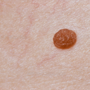 Compound Nevi
- Compound nevi are usually raised and skin-colored, with uniform pigmentation.
- These moles have features of both junctional and intradermal nevi.
- They are acquired nevi.
- The melanocytes that make them are located both in the dermo-epidermal junction and the dermis.
 Acquired Nevi
- Acquired nevi appear during childhood or adulthood.
- They are benign (non-cancerous) lesions.
- Although it is rare, these moles do have the potential to change into melanoma.
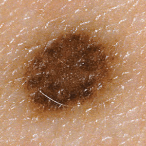
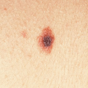
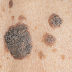
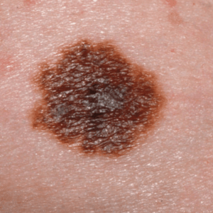 Atypical Nevi (Dysplastic Nevi)
- Atypical nevi are not ordinary moles; they are generally larger, with irregular or indistinct borders and variations of color within the mole, ranging in color from pink to dark brown.
- Sometimes they are described as looking like fried eggs, with a raised center of darker pigmentation surrounded by a flat, lighter area.
- Atypical nevi may occur anywhere on the body but are found most frequently on the back and on sun-exposed areas.
- The vast majority of atypical nevi do not become melanomas, but having atypical nevi is a risk factor for melanoma.
- Familial Atypical Mole and Melanoma (FAMM) syndrome, also called dysplastic nevus syndrome, is an inherited condition in which affected individuals develop many moles (usually at least 50 to 100) and their moles are often atypical
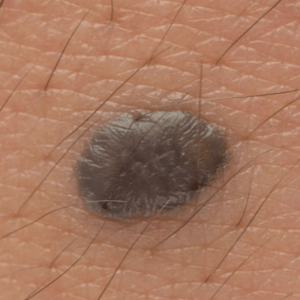 Blue Nevi
- Blue nevi are blue-gray to blue-black moles that are present at birth or appear later in life.
- They are categorized into either a “common blue nevus” or a “cellular blue nevus.” The common blue nevus is typically flat or dome-shaped with a smooth surface and ranges in size from a 0.5-1cm. Cellular blue nevi are at least 1 cm in diameter and can grow in size over time and the surface can ulcerate.
- These nevi are most commonly found on the head and neck, sacral area (above the tailbone), back of hands, and feet. However, they can be found on other areas of the body.
- It is unknown whether there is a genetic component, but they are frequently found in Asian populations and more often on women than on men.
- Most blue nevi are benign although there is a possibility that a cellular blue nevus can become cancerous. Therefore, if you have a changing or suspicious lesion, you should bring the nevus to the attention of your healthcare provider.
 Congenital Nevi
- Congenital nevi are moles that you are born with or that appear in early infancy. They are uncommon, appearing in only about 1% to 2% of all newborns[1-3]
- The risk of developing a melanoma is directly related to the size of the congenital nevus.
- Studies that have investigated the risk of a small or medium congenital nevus turning into a melanoma have put the lifetime risk at between 0% – 5%. These nevi are tan in color and flat or mildly palpable. However, over time, they become darker and raised.
- The risk for giant congenital nevi (also known as garment nevi), which cover major areas of the head or body, is less clear. Historically, some studies reported a very high lifetime risk, up to 50%. Recently, larger studies have placed the lifetime risk at closer to 10% – 12%.[4] These nevi can have irregular color patterns and are often accompanied by multiple smaller “satellite” nevi.
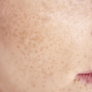 Freckles
- Freckles are small, tan, flat spots that appear in response to the sun’s ultraviolet radiation.
- The medical name for a freckle is an “ephelide.”
- Freckles darken with sun exposure and lighten or fade when no longer exposed. They tend to cluster together in large numbers.
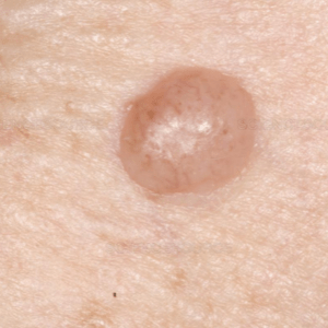 Intradermal Nevi
- Intradermal nevi are flesh-colored or light brown dome-shaped lesions.
- Another name for these moles is “dermal nevi.”
- The melanocytes that make up an intradermal nevus are located in the dermis (below the dermo-epidermal junction). This mass of melanocytes pushes the cells above it upward, resulting in the flesh-colored bump that is noted on the surface of the skin.
- They are acquired nevi most commonly found in adults.
- Nonpigmented (not colored) dermal nevi are also called “cellular nevi.”
 Junctional Melanocytic Nevi
- Junctional melanocytic nevi are acquired nevi that tend to first appear in childhood as flat, freckle-like lesions of brown, dark brown, or black, and are uniform in color.
- These moles are called “junctional nevi” because the melanocytes that make them are located at the junction of the epidermis and dermis.
- They are most commonly found on the face, arms, legs, trunk, genitals, or soles of the feet.
- During adulthood, these nevi may become more raised, lose pigmentation, or even disappear.
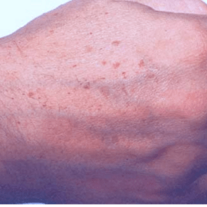 Lentigines
- Lentigines (singular: lentigo) are flat, brown, sometimes large spots that are associated with sun-damaged skin.
- They are also known as sunspots, age spots, or liver spots, although they have nothing to do with the liver or liver function.
- Unlike freckles, they are usually discrete spots that do not fade when no longer exposed to the sun.
- Lentigines are very common in people over 40.
- You’ll find them in areas of greatest sun exposure: on the face, arms, chest, shoulders, back, and backs of the hands.
- IMPORTANT! Because lentigines are a marker for sun damage, people with lentigines are at increased risk for developing melanoma.
 Seborrheic Keratoses
- Seborrheic keratoses are raised, benign growths of keratinocytes in the upper layers of the skin (the epidermis).
- These keratinocytes are often pigmented, which gives the seborrheic keratosis its dark, mole-like appearance.
- They are sometimes mistaken for warts or melanoma and range in color from tan to dark brown or black. They have a waxy, pasted-on appearance which is why they are sometimes described as “brown candle wax on the skin.”
- These lesions can appear on sun-exposed or covered areas. They never grow on the palms or soles or in the mouth or eyes.
- Unlike moles, seborrheic keratoses are usually rough to the touch.
- Seborrheic keratoses are the most common benign lesions that mimic the appearance of melanoma. Although a seborrheic keratosis cannot turn into a melanoma because it is not made of melanocytes, it cannot be completely ignored because melanoma can occur next to them and may be mistaken for an extension of the seborrheic keratosis.
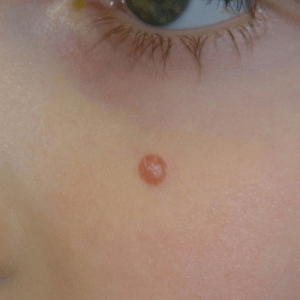 Spitz Nevi
- Spitz nevi often have features that overlap with those of melanoma, but Spitz nevi are benign and melanoma is, of course, cancerous.
- They are most often a pink, raised bump, but they are sometimes red, blue, or black and even non-pigmented.
- These lesions typically develop on the face, neck, and legs but can also be found on the arm, shoulders, or trunk. They grow rapidly and can be 1 cm in size within six months. After that time, they should remain the same size, shape, and color. After several years, they may even get smaller or disappear.
- They most frequently develop on fair skinned individuals and are more common among a younger population.