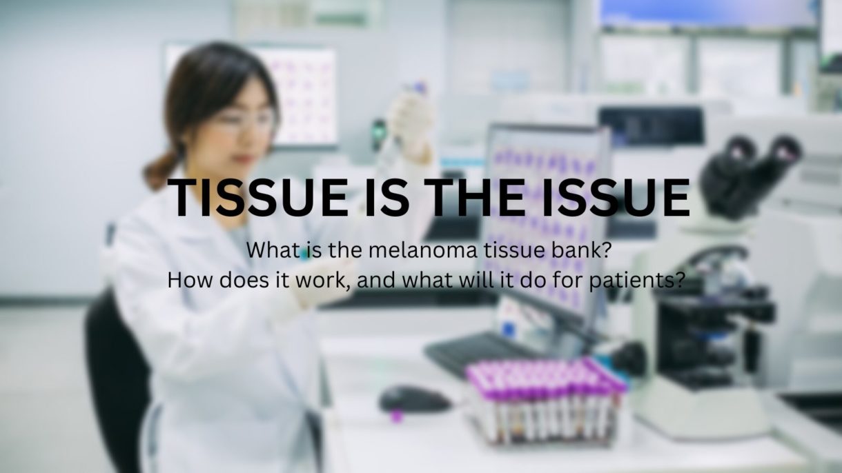
TISSUE IS THE ISSUE—2023 Update on AIM’s International Melanoma Tissue Bank Consortium
Published:
01/26/2023
This article is an update of an article we published last year.
AIM at Melanoma’s major research initiative—the International Melanoma Tissue Bank Consortium (IMTBC)—is the first of its kind in the world. Currently comprising four sites in the U.S. (with two Australian sites to be added), the IMTBC is a resource of primary melanoma tumor tissues and data for use by the participating institutions’ researchers as well as researchers around the world who can apply to use the tissue/data.
As we noted last year, COVID hit research projects like this hard, as only “essential” medical appointments were scheduled during the pandemic. And since then, offices and patients have been slowly getting back to normal. As you’ll read below, the typical way we collect a primary tumor tissue is at one of four research institutions during a skin check appointment. Since these institutions are also major hospitals, getting back to normal has been complicated because COVID affected hospitals in a much different way than it did independent physician offices. But we are moving in the right direction, and collection resumed and continued at all four U.S. sites in 2022. As of the end of the 3rd quarter (September 30, 2022) reporting, we have collected 110 fresh frozen primary tissues and corresponding data.
The following is an FAQ about the tissue bank. Some of the questions we’ve published previously; some are new.
What is the tissue bank—is it an actual building?
The IMTBC is not a building. It operates within certain existing medical centers. Dermatologists, surgeons, oncologists, pathologists, and other staff all participate in creating the bank at a participating cancer center.
How does it work?
Here’s how it typically works: A patient goes to see his or her dermatologist for a skin check at one of the tissue bank locations. The physician finds a lesion suspicious for melanoma and intends to biopsy it. Before the excision begins, the physician asks for consent from the patient to use some of the tissue and the patient’s medical information—depersonalized—for research. If granted, a portion of the tissue is removed for the biopsy, and a portion is frozen immediately, which, critically, preserves RNA. The doctor also asks the patient to fill out a special questionnaire and collects other samples such as blood. Staff enter the patient’s medical record and questionnaire answers (again, all depersonalized) into a special database, and the samples and tissue are coded and stored in refrigerators/freezers.
The bank, then, is made up of both fresh frozen tumor tissue and other samples, such as blood, as well as data about each patient.
What will the researchers do with the tissue and accompanying patient information?
Once a critical mass of tissues and data are collected from a variety of patients, the researchers can look at these fresh frozen primary melanomas and the accompanying data to find medical signs or indications, called biomarkers, that are shared by some or all of the tissues. These biomarkers will help reveal insights into diagnosis, prognosis, and treatment.
Please read this article for even more information.
Can I participate?
This question is the most common one we get.
At this time, only certain people can participate, and they likely won’t be anyone who is reading this article. Why? Remember that we are collecting primary melanoma tumor tissue—primary means the original melanoma found on your body, the one that was deemed Stage I or Stage II. Those who are reading an article like this are probably long past that original biopsy—though they may be able to participate if they have a second primary melanoma. Further, the patient needs to be seen at one of our tissue bank sites at Hillman Cancer Center, University of Pittsburgh Medical Center (Pittsburgh, PA); Knight Cancer Institute, Oregon Health and Science University (Portland, OR); California Pacific Medical Center (San Francisco, CA); or Robert H. Lurie Comprehensive Cancer Center, Northwestern University (Chicago, IL). Finally, the suspected melanoma must be large enough to bank.
So ironically, it is people likely unfamiliar with the melanoma world and melanoma research—because at the time of the biopsy, they won’t even know if they have melanoma—whose information and samples will populate this bank and help researchers understand melanoma better.
We hope in the future to have more U.S. sites so we can collect more primary tissue, but for now, we are limited to collecting at the above four U.S. locations.
Why is IMTBC unique?
IMTBC is a global first because of the following combination of factors:
- It’s a consortium—the investigators at the institutions are sharing data and tissue samples with each other
- It’s collaborative—tissue samples and data will be available for researchers around the world to apply to study
- The tissue is fresh frozen—RNA is preserved, unlike in the standard formalin fixed, paraffin embedded process
- The tumors are primary—not metastasized
- There will be a critical mass—a goal of 500 to start, and continued collection thereafter
- Full annotation will accompany each tissue—patient data, including full medical history (depersonalized), will be available for study along with the tissue
- Samples accompany each tissue—blood and other samples are collected for each patient
Why is fresh frozen tissue important?
Formalin-fixed, paraffin imbedded is the standard way of storing primary tumors. In this method, a lot of water is depleted from the tissue, which degrades the proteins. The messenger RNA is also degraded into smaller pieces during the fixing process, and researchers want the messenger RNA intact. Fresh frozen tissue preserves RNA. Finally, a tumor cell has already initiated an immune response, and researchers are able to recognize that in a fresh frozen piece but not in a formalin-fixed piece.
Why is primary tumor tissue important?
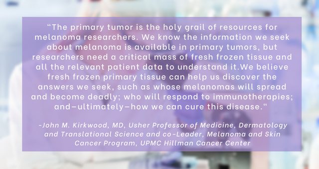
The information in the primary tumor is important because it is fixed. The heterogeneity of the disease can only be read in the primary tumor—it’s the best place to understand the heterogeneity of the disease. There is a tremendous amount of information in the primary tumor that can help us predict the future for the patient if we can learn to read it. The primary tumor has ALL the information about the disease.
The highest degree of genetic alterations is found in the primary tumor. During the course of the disease, the tumor loses large DNA fragments. For research, you want to look at the tumor at several “time points,” if possible, starting most importantly with the primary tumor.
If we are going to find predictive or prognostic markers, it will be in the primary tumor.
By contrast, metastasized tumors are a product of the organ in which they are growing, and they will reflect that organ. Some tumor cells go to the lymph nodes, and other cells go into the bloodstream, so you don’t get a full picture of the person’s disease from the metastases.
If primary melanoma tissue that is fresh frozen is so important for research, why isn’t there more available?
One reason is that fresh frozen primary melanoma tissue cannot be collected and frozen in a typical dermatologist’s office, and a typical dermatologist’s office is where most melanoma biopsies take place. Most dermatology offices do not have an in-house pathologist, and therefore the dermatologist must physically send their biopsies to an outside pathology office. That situation eliminates the ability to safely separate a portion of the sample and flash freeze it for research without compromising the diagnosis, particularly if the melanoma is small. Even when a dermatologist’s office has an in-house pathologist, most dermatology/pathology offices do not have the time, set-up, or all of the materials needed to quickly freeze the tissue (using liquid nitrogen) or store it (liquid nitrogen freezers operate at –140°C to –196°C). Part of the samples also need to be processed immediately, requiring an on-site laboratory, which is not available in a typical dermatology clinic. And none of the above difficulties includes the very complicated procedures and rules around gathering consent of patients for tissue collection and using that tissue for research—again, a typical dermatology office does not work in this realm.
Dermatology offices located at research institutions, however, with on-site pathology offices and on site laboratories and appropriate space and equipment, are the places that can successfully separate, freeze, and store fresh frozen primary tissue for research. But even these places need to be organized, managed, and paid to consent patients appropriately; to put the collection processes in place; to label and store tissue; and to oversee the use of the tissue by internal and external researchers. This organization, management, and funding are what IMTBC has successfully accomplished.
What happens next?
Critical mass is important. We are at 110 tissues, and the site researchers wanted 100 tissues at the very least to begin their research—the more tissues, the more that can be revealed through their studies—so this year, we should see the first research project(s) using the tissue.
We are in initial conversation with another U.S. institution to join the consortium, and we are in continued conversation with our Australian sites on the “how” of collection, collaboration, and processes and hope that they can officially open soon. We will announce any and all news as it happens!
Recent Posts

Jul. 20, 2026
More Than a Letter: A Year of Advocacy for Patients with Advanced Melanoma

Jul. 14, 2026
Finding Hope, Purpose, and Life Beyond Diagnosis

Jul. 10, 2026
Help Bring a New Treatment Option to Patients with Advanced Melanoma

Jul. 06, 2026
How Melanoma Changed Adam Harper’s Perspective on Life

Jun. 26, 2026