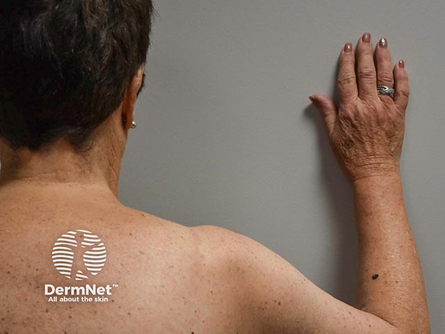
What Does Melanoma Look Like?

Melanoma: Recognize It Early. Act Quickly.
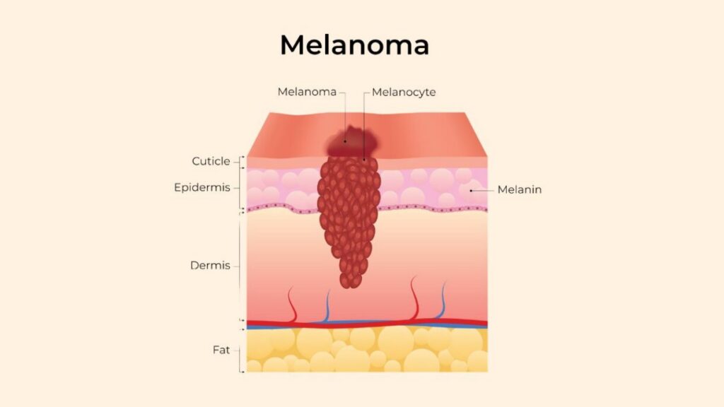
Melanoma, a type of skin cancer, is one of the most aggressive and deadliest forms of skin cancer. It develops when the melanocytes, the cells responsible for producing melanin, which gives color to our skin, hair, and eyes — mutate and become cancerous. Identifying melanoma at an early stage is crucial for successful treatment. Therefore, it is important to understand what melanoma looks like.
While these images may provide some general information about the appearance of melanoma, they cannot accurately depict every possible variation. Therefore, if you notice any suspicious moles or marks on your body, do not hesitate – seek medical attention promptly and prioritize your health over internet comparisons.
The earlier melanoma is diagnosed by a qualified healthcare professional, the easier it becomes to treat effectively. This information does not constitute medical advice or diagnosis.
NOTE: Relying solely on online images is not advisable and can be potentially dangerous.
All images sourced from DermNet
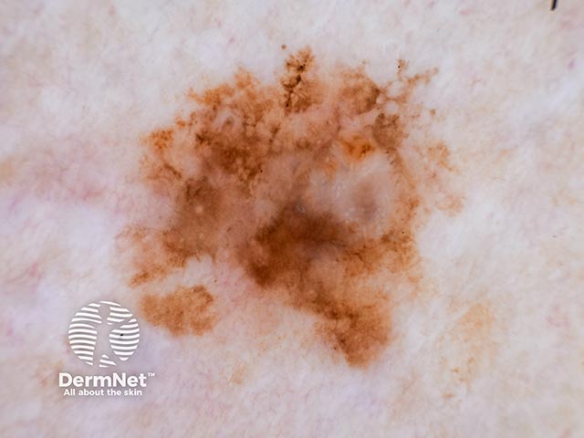
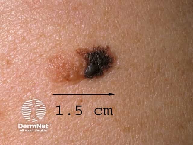
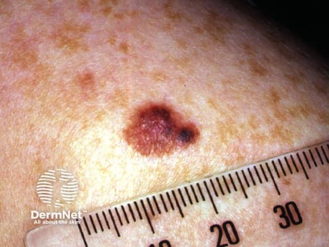
Cutaneous Melanoma Images
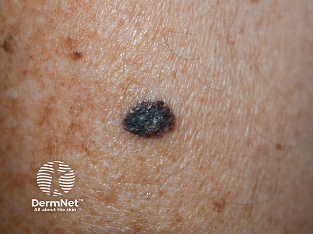
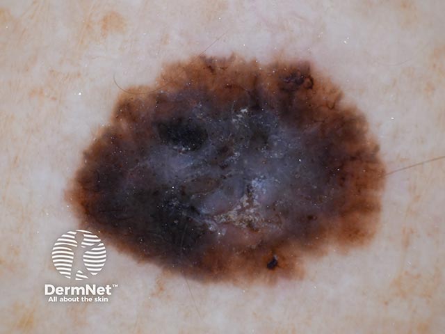
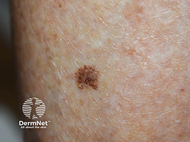
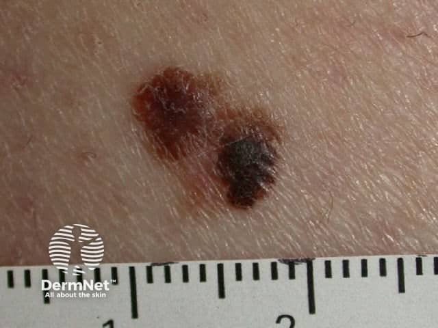
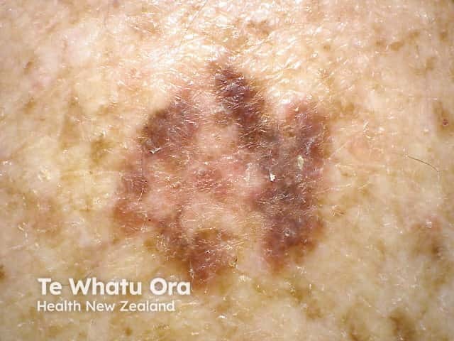
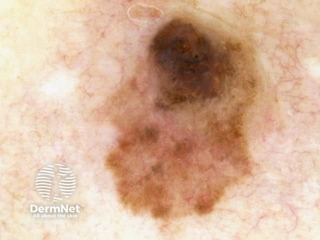
Superficial Spreading Melanoma
Superficial spreading melanoma (SSM) is the most common subtype of melanoma.
- It is a type of cutaneous melanoma that typically begins in the epidermis (the outer layer of the skin).
- It is characterized by a prolonged horizontal (radial) growth phase, meaning it spreads across the surface of the skin before growing deeper.
- This early growth pattern often makes it more detectable at an earlier stage compared to more aggressive subtypes.
What are the signs of superficial spreading melanoma?
- Often appears as a flat or slightly raised lesion
- Has irregular borders and multiple colors (such as tan, brown, black, red, or white)
- Commonly develops on sun exposed areas, such as the chest, back, legs
- Can arise from an existing mole or appear as a new lesion
Biological behavior
- During the radial growth phase, the melanoma cells spread laterally within the epidermis.
- Over time, it may enter a vertical growth phase, where it invades deeper layers of the skin, increasing the risk of metastasis.
Importance
Because superficial spreading melanoma grows outward before downward, early detection significantly improves outcomes, making skin checks and monitoring changes critical.

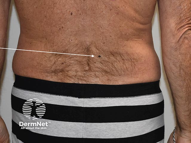
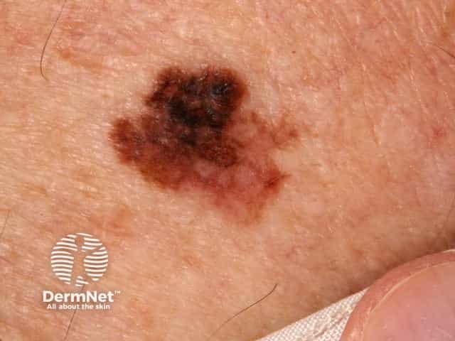

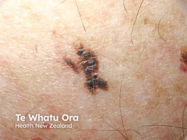
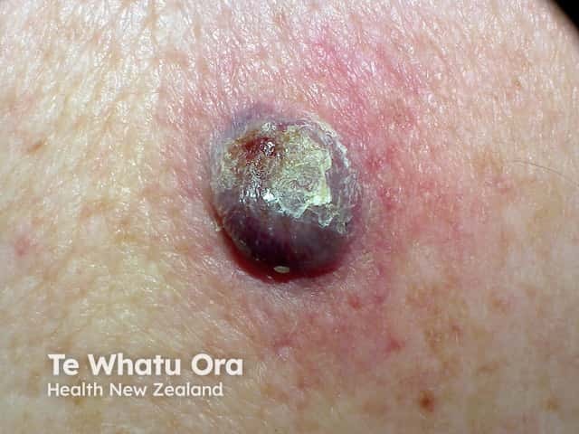
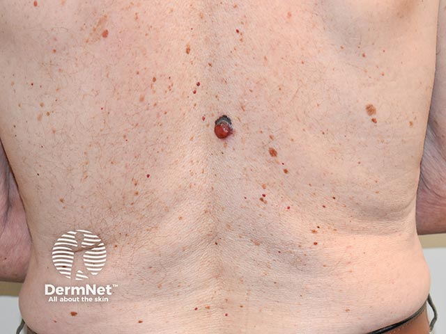
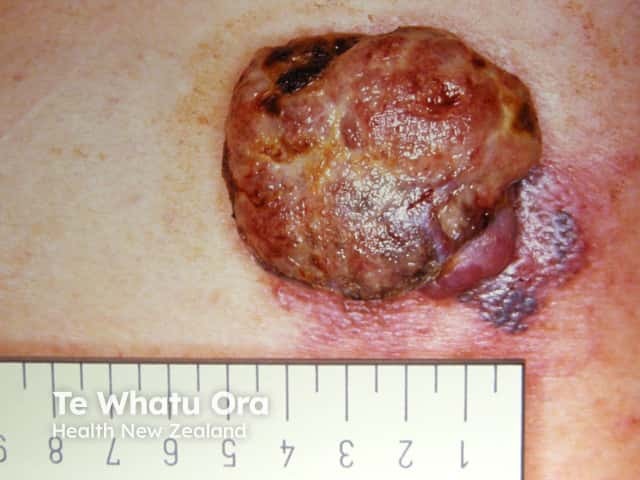
Nodular Melanoma
Nodular melanoma is a type of cutaneous melanoma and the second most common subtype
- It is characterized by its aggressive behavior and rapid growth
- It accounts for 10% to 15% of all cases of melanoma
- It is often diagnosed at a more advanced stage, which can make treatment more challenging
What are the signs of nodular melanoma?
- Typically appears as a raised, dome-shaped bump
- May be red, pink, brown, or black in color
- Often lacks the classic warning signs (asymmetry, border irregularity, color variation)
- Most commonly found on sun-exposed areas, especially the head and neck
- Can develop anywhere on the body
Biological behavior
- Nodular melanoma often enters the vertical growth phase immediately
- This means it grows downward into deeper layers of the skin early, increasing the risk of metastasis
- Rapid growth is the most important clinical clue
Importance
Nodular melanoma can be harder to recognize because it does not always follow typical melanoma warning signs. A key indicator is anything on the skin that is growing quickly and looks different from other spots. Early evaluation by a dermatologist is critical, as early detection greatly improves treatment outcomes.
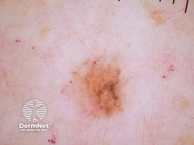
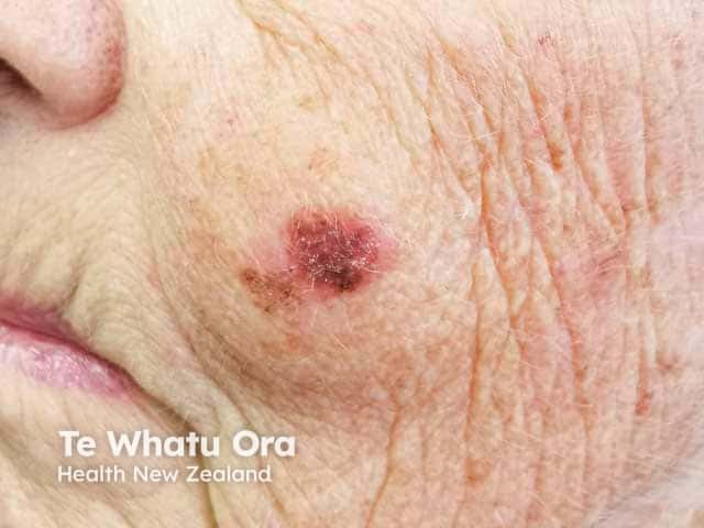
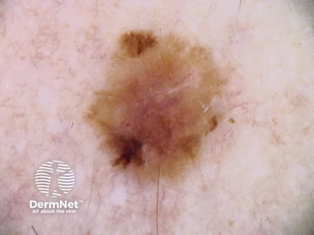
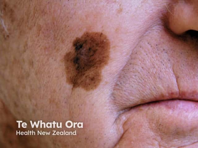
Lentigo Maligna Melanoma (LMM)
Lentigo maligna melanoma (LMM) is a subtype of cutaneous melanoma.
- It originates from melanocytes, the cells that produce melanin (skin pigment)
- It most commonly occurs in older individuals with a history of chronic sun exposure
- It typically develops on sun-damaged skin, especially the face and hands
What are the signs of lentigo maligna melanoma?
- Appears as a flat lesion with an irregular shape
- Shows uneven coloration, often with varying shades of brown or black
- Commonly found on areas with long-term sun exposure
- Can resemble benign lesions such as freckles or age spots, making recognition more difficult
Biological behavior
- Begins as lentigo maligna, a slow-growing, in situ (non-invasive) phase
- May eventually progress into lentigo maligna melanoma, where it becomes invasive
- Once invasive, it has the potential to spread to other parts of the body
Importance
Because lentigo maligna melanoma can closely resemble harmless skin markings, early detection can be challenging but is critical. Identifying changes in sun-damaged skin and seeking evaluation can help catch it before it becomes invasive, when treatment is most effective.

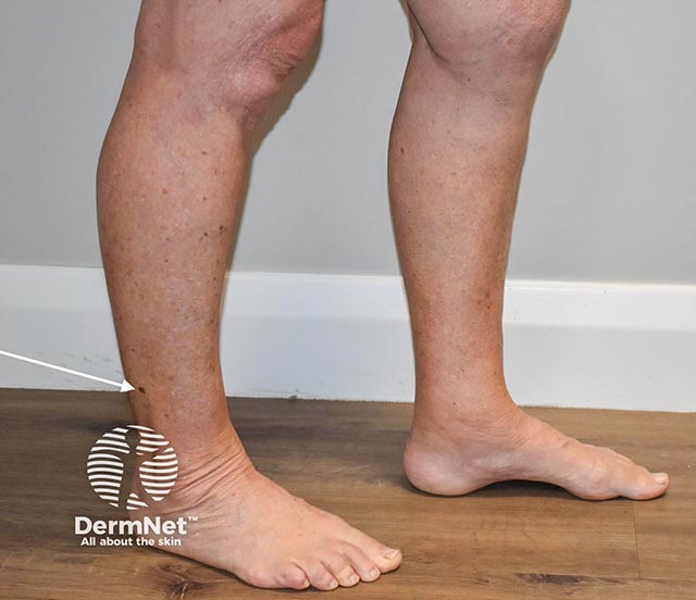
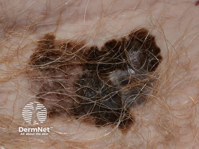
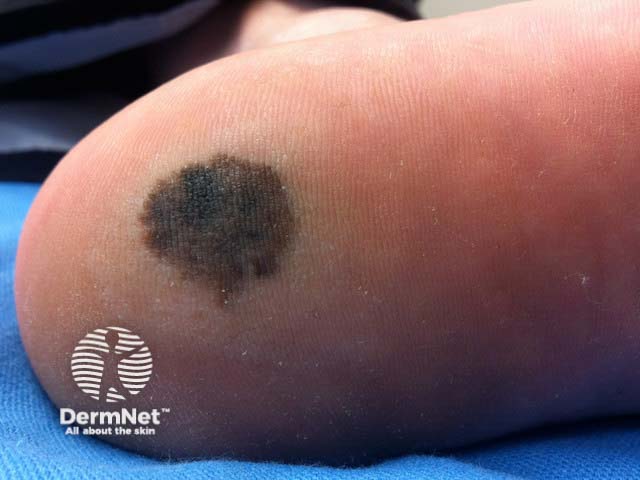
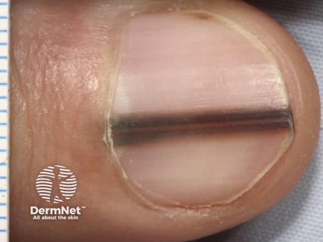
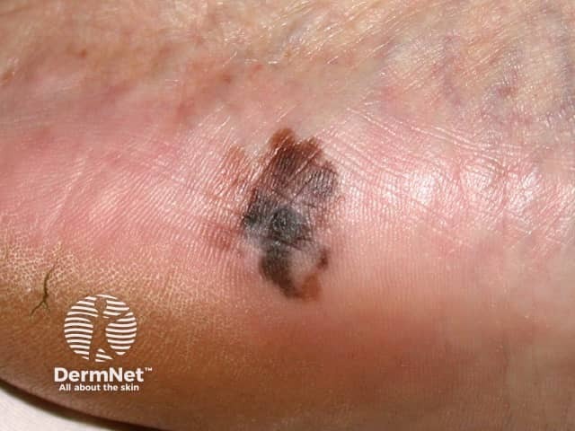
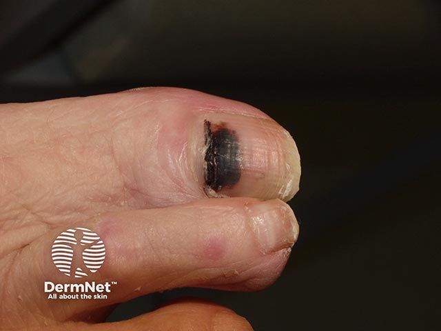
Acral Lentiginous Melanoma (ALM)
Acral melanoma is a rare subtype of cutaneous melanoma.
- It primarily affects the palms of the hands, soles of the feet, and under the nails
- Unlike many other melanomas, it is not strongly associated with sun exposure
- Because it occurs in less visible areas, it can be more difficult to detect early
What are the signs of acral lentiginous melanoma?
- Appears as a dark spot or patch on the palms or soles
- May present as a new or changing growth under the nail (subungual melanoma)
- Can be mistaken for bruising, stains, or other benign conditions
- Often develops in areas not routinely examined
Biological behavior
- May go unnoticed in early stages due to its location and subtle appearance
- Delayed detection can allow for progression to more advanced disease
- Like other melanomas, it can become invasive and spread if not identified early
Importance
Any new or unusual lesion on the palms, soles, or under the nails should be evaluated promptly. Early recognition is critical, as timely diagnosis significantly improves treatment outcomes.
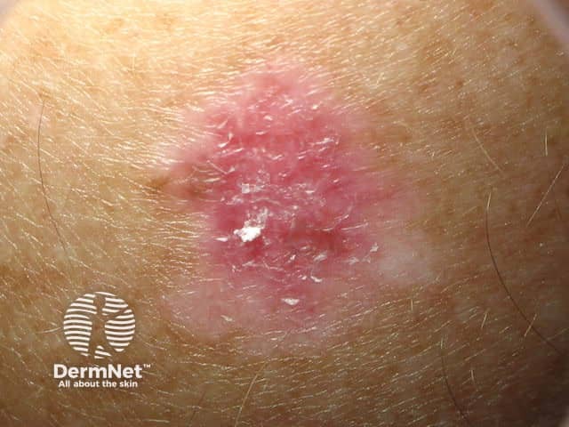
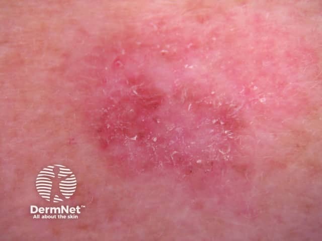
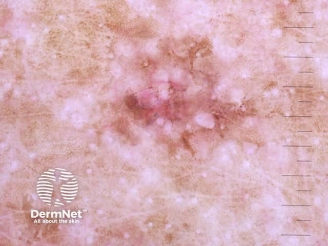
Amelanotic Melanoma
Amelanotic melanoma (AM) is a rare subtype of cutaneous melanoma.
- It is characterized by a lack of melanin (pigment), which makes it more difficult to recognize
- It is considered aggressive and is often diagnosed later than more typical melanomas
- The absence of pigment means it may not resemble a typical dark melanoma lesion
What are the signs of amelanotic melanoma?
- Often appears as a pink, red, or skin-colored lesion
- May present as a nodule, patch, or ulcer
- Can be mistaken for benign skin conditions such as eczema or psoriasis
- Lacks the dark coloration commonly associated with melanoma
Biological behavior
- Because it is harder to identify visually, diagnosis is often delayed
- Delayed detection can allow for progression to more advanced stages
- Like other melanomas, it can become invasive and spread if not treated early
Importance
Amelanotic melanoma can be easily overlooked due to its atypical appearance. Any new, changing, or unusual skin lesion—especially one that does not heal—should be evaluated promptly, as early detection is critical for improving outcomes.