Sentinel Lymph Node Biopsy
What Is A Sentinel Lymph Node Biopsy?
Once you have been diagnosed with melanoma, the next step is to assess your risk of spread beyond the primary tumor.
If your doctor believes the risk is high enough, s/he’ll perform further tests to establish whether the melanoma has spread. One of those tests is sentinel lymph node biopsy.
The presence or absence of melanoma cells in the lymph nodes is one of the most important prognostic factors we have, since it indicates a high risk that the melanoma will spread to distant sites in the body.
Your doctor will feel your lymph nodes during a physical exam. If any nodes feel enlarged, irregular, or firm, it may be an indication that the melanoma has spread. If they are enlarged, irregular, or firm, your doctor may recommend a fine needle aspiration biopsy and not a sentinel lymph node biopsy.
The Role of The Sentinel Lymph Node Biopsy (SLNB)
SLNB is a specialized procedure performed to determine whether any melanoma cells have spread to the lymph nodes. If the melanoma has spread, it will usually spread to the lymph nodes nearest the area of the primary melanoma. The sentinel lymph nodes are the first of those lymph nodes to receive drainage from the primary tumor, and therefore the ones most likely to have melanoma cells if any of them have spread.
SLNB is generally considered when one or more of the following are true:
- The melanoma is equal to or greater than 1.0mm in depth
- There is an ulcerated tumor of any thickness
- The margins taken during the biopsy are positive for melanoma
- There is lymphovascular invasion (seeing cancer cells in the lymphatic channels or blood vessels)
SLNB is generally not considered if:
- The melanoma is less than 0.76mm with no other risk features
- It is already known that melanoma is in the lymph nodes (Stage III)
- The melanoma has spread to distant organs (Stage IV)
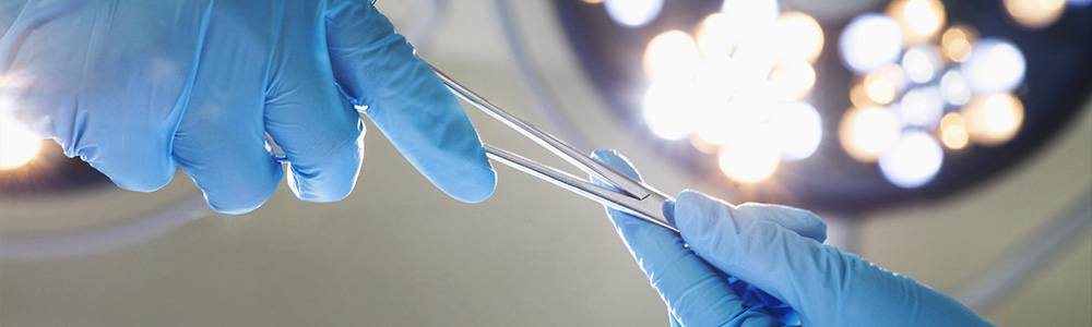
How SLNB is Performed
The SLNB has two parts, a radiology test called lymphatic mapping and a surgical procedure. A wide local excision is generally performed at the same time.
Lymphatic Mapping (Lymphoscintigram) usually involves injecting radioactive dye in the skin around the site of the original melanoma. Then a special camera is used to watch the radioactive material move from where the melanoma was biopsied to the group of lymph nodes the melanoma is most likely to travel to first. These are called the sentinel nodes, and most patients have between one and five sentinel nodes. Surgery is performed after the lymphatic mapping has been completed. You will generally receive a second agent (blue dye) that will help visually identify the lymph nodes that have already been detected using the special camera. This two-method approach is more accurate than using either one alone. The surgeon will remove the sentinel nodes and send them to a pathologist or dermatopathologist for review.
Wide Local Excision is a procedure in which the melanoma—including the biopsy site and an area of normal tissue around it (margins)—is removed. It is recommended that lymphatic mapping and the lymph node surgery be performed before the wide local excision is done.
After SLNB
The sentinel nodes removed by your surgeon will be examined under a microscope by a pathologist or dermatopathologist to determine if there is melanoma in them. If the sentinel lymph nodes do not show evidence of melanoma, it is unlikely that the cancer has spread to the remaining lymph nodes and no further surgery is necessary. If one or more of the sentinel lymph nodes is positive for melanoma, the remaining lymph nodes in that area may be removed. This procedure is called a completion lymph node dissection (CLND). Your doctor will discuss the need for CLND with you, and your treatment type may affect whether you have CLND.
Note: The survival rate of patients is markedly better when the melanoma in the lymph nodes has been found by means of a SLNB, as opposed to being found during a physical exam.